What is MND
Find support
I have MND
I am supporting someone
Get involved
Research
About MND Scotland
What’s new?
© MND Scotland 2025
© MND Scotland 2025

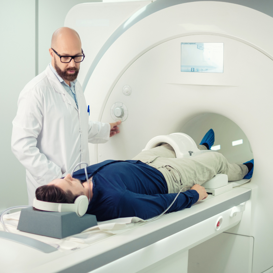
The new research project, run by Professor Roger Whittaker, a consultant clinical neurophysiologist at the University of Newcastle, will investigate whether it is possible to lay the foundations for a non-invasive investigation using standard hospital MRI scanners.
MND is characterized by a progressive loss of muscle function which results from the loss of specialist cells, motor neurons, which control the muscles. Motor neurons are vital for activities such as moving, speaking, eating and breathing. A single motor neuron controls a number of muscle fibres, and this is called a motor unit. One of the earliest symptoms of MND is often fasciculations, which are involuntary contractions in muscles, caused by faulty motor units in those affected by MND.
Professor Whittaker and his team have developed a technique which allows them to detect the activity of individual motor units using an MRI scanner, which they call motor unit MRI (MUMRI). When a motor unit contracts it appears as a darker area on the MUMRI image and, in those who experience fasciculations, these motor units can be identified even in a resting state. This technique has already been trialled, using a routine MRI scanner, in a small number of patients in the NHS trust in Newcastle.
Although fasciculation is an important early sign of motor unit dysfunction, it is not enough on its own to make a diagnosis of MND. MND Scotland is providing £225,000 funding over three years to further develop this work so that additional motor unit changes – not just fasciculations – can also be imaged and assessed in a single scan session. This will be achieved by imaging the motor units after initiating a gentle muscle contraction.
Currently MND diagnosis is a very time-consuming process and can require the individual to undergo invasive and painful tests. This new approach may make it possible to not only diagnose MND in an entirely non-invasive and pain-free way, but also track the progress of the disease over time, since the rate of motor unit degradation can be monitored. This is crucial for developing new treatments as it would allow researchers to quickly identify those treatments that are working, and switch patients away from those that are not.
Over the first year, the team of experts based at the University of Newcastle, will focus on refining the scan technique. They will then aim to invite a group of MND patients to test it, requiring them to undergo a MUMRI scan every six months for 18 months. These regular scans will determine whether MUMRI performs better than current diagnostic and disease progression tracking methods.
This project will be a real team effort, involving clinical neurophysiologists, neuroradiologists, MR physicists, computer scientists and trial statisticians. Joining Professor Whittaker’s team to undertake this project for MND Scotland will be Dr Ao Wang. Ao completed his PhD at York University using MRI to study brain activity, and this project is an ideal opportunity for him to use these skills in a new area.
Ao said: “This will push my research from theoretical and abstract to real life. This triggers my passion; I could not stop thinking about the future if a new and better biomarker for MND progression could be applied to all patients in need.”
Professor Whittaker said; “I’m really excited about this project and am delighted to have Ao joining the team. We desperately need faster and more accurate ways to diagnose and monitor people with MND. If this project is successful, it opens the door to achieving this with a single MRI scan. There is a lot of work still to do, but with this generous funding from MND Scotland we can get started right away.”
Dr Jane Haley, MND Scotland’s Director of Research said “Our 2022 “MND What Matters” survey identified the length of time it takes to get a definitive diagnosis is an area of real concern for people with MND. While many factors contribute to this delay, the lack of a clear diagnostic tool is a significant issue.
“We hope the work that Professor Whittaker and his team are undertaking will lead to the development of a diagnostic test that can be administered painlessly in any hospital with an MRI scanner. If this can also be used as a biomarker for monitoring disease progression in clinical trials, that will be an additional, and important, benefit.”

Sign up
for newsletter
Get the latest news and events straight to your inbox.
You can help create a world without MND