What is MND
Find support
I have MND
I am supporting someone
Get involved
Research
About MND Scotland
What’s new?
© MND Scotland 2026
© MND Scotland 2026
Dr Chris Henstridge is a leading neuroscience researcher based at the University of Dundee who has a wealth of experience in researching MND. Chris has studied the connections in our brain and how, in neurodegenerative diseases, neurons are damaged and the variety of symptoms this can cause. He was a key part of setting up the world’s first human MND brain bank, helping to propel MND research forward. Chris has been involved with MND Scotland for many years as a researcher, has been involved in our LEARN events from the beginning, and has even taken part in fundraising efforts.
In 2021 MND Scotland and Alzheimer Research UK funded £61,196 to Dr Francisco Inesta Vaquera and Dr Chris Henstridge to research preclinical animal models of MND to speed up drug discovery.
We at MND Scotland understand the importance of animal models in research, and aim to uphold our values of reduction, replacement, and refinement within research. Animal models are used when necessary, with no adequate alternative. Please read our statement on animal use in research.
Now that the project has finished, we asked Chris to explain what an animal model is, their importance in MND research, and how their new animal model will affect future MND research.
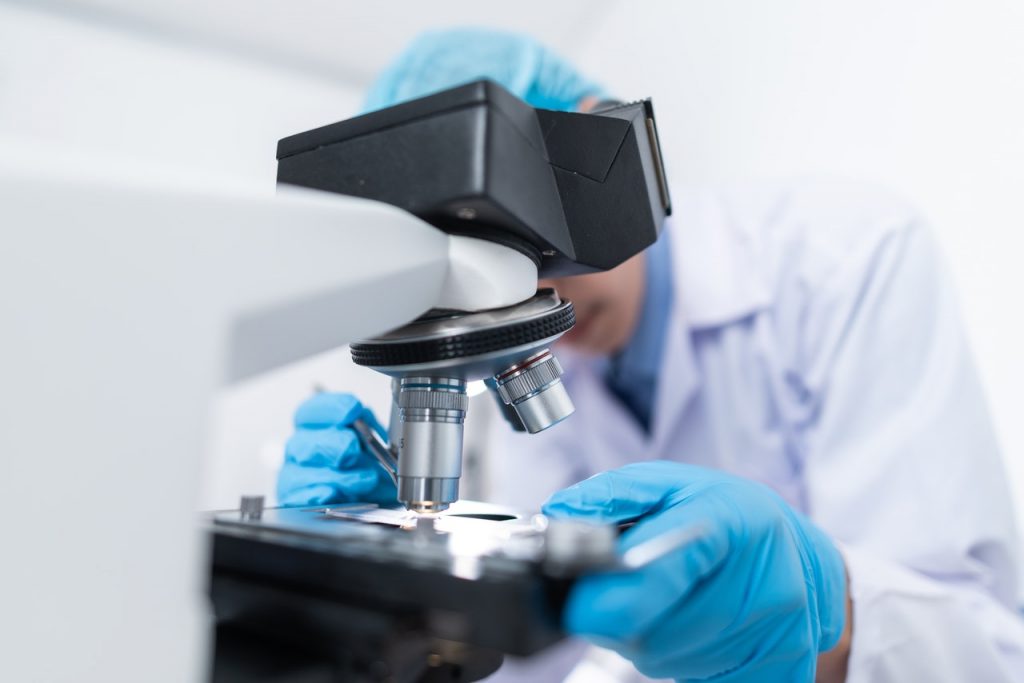
My name is Chris, and I’m a senior lecturer at the University of Dundee. I have always been interested in the connection points between brain cells, called synapses, and have studied how they change in disease for around 15yrs. I became involved in MND research when I was a PostDoc in Edinburgh, working closely with the Edinburgh Brain Bank. I realised there was a unique opportunity to look at donated human tissue to study changes in the synapse to explain some of the symptoms of MND. I was particularly focused on finding a link between changes in thinking skills and loss of synapses. The results from that early work sparked my career in MND research and my team in Dundee are now focused on discovering why synapses break down in MND and thinking of ways to stop it.
Our human work is essential to understand what the disease does to the brain and spinal cord of people living with MND. However, we cannot take samples of the brain during life like we can with liver or skin for example, so the brains we receive can only show us the end stage of the disease. To understand the early steps in MND, we must use models.
An animal model is a non-human species that we use to mimic a certain biological process or disease, like MND. In our work we use mice that have had certain aspects of their DNA altered to characterise a certain aspect of MND.
Lots of amazing work has been done in cells grown in a dish, however in most cases these cells grow in a single layer on the bottom of the dish. This does not model our brain very well, so other teams use intact animal models instead. An intact animal brain contains many of the different cell types and connections that we have in our brain, so is a better model to help understand how MND starts and spreads through the body. However, it is essential to understand that these are just models and must be used wisely. They can help us answer very specific questions but cannot give us the whole answer. This is why lots of different models are used by researchers all over the world. Induced pluripotent stem cell (IPSC) technology has revolutionised research, allowing research teams to collect skin cells from people during life and turn them into brain cells that can be grown in the lab. This means researchers can now study human brain cells in a dish from people living with MND.
There are lots of different types of animal models for MND, including in fish and rodents. A few of occur naturally although most are created, and these allow researchers to undertake studies to increase our understanding of the various mechanisms that might underpin MND. One example is the research to understand the involvement of a gene called SOD1 (superoxide dismutase 1). One form of MND is associated with multiple changes in the SOD1 gene – over 100 different changes have been identified to date, though not all are associated with the disease. This discovery in the 1990’s meant that researchers were hopeful that they could develop rodent models that might mimic the human condition. Since then, more than 10 different rodent models have been developed with alterations in the SOD1 gene and these have allowed researchers to ask questions and investigate disease changes, that would not be possible in people or in cells in a dish. At least one of these rodent models was used during the early-stage development of tofersen, a new targeted gene therapy treatment for SOD1 MND that received regulatory approval in the USA and Europe in 2023.
In MND a protein called TDP-43 begins to malfunction and moves to parts of a cell it isn’t normally found in. Then it clumps together and stops the cells from working properly, leading to cell stress. This triggers lots of different changes in the brain and other parts of the body. However, we don’t know exactly where or when cells become stressed, so we designed a model system to help us find out. Our mouse model was carefully designed to add a flag to cells that were becoming stressed due to TDP-43 malfunction. We can now look to see where and when these flags appear, which can give us important clues about the earliest changes in MND.
This new model can be used in several ways. Firstly, it can help show us where and when cells first become stressed in MND. Secondly, it can help us discover which cells are most likely to be damaged in MND, and thirdly it could be used as a tool to discover if new MND drugs are effective by reducing cell stress.
This new model was designed by Dr. Francisco Inesta-Vaquera, Prof. Roland Wolff and I in Dundee, and is ready for researchers to use. We have performed some initial characterisation and revealed a vulnerable cell type in the brain that becomes flagged early in MND. We hope that teams in universities and drug companies around the world will use this to discover new understanding of MND and develop ways of treating it.
MND Scotland would like to congratulate Dr Chris Henstridge and Dr Francisco Inesta Vaquera on their success in making a new model for research use. Projects like this allow for greater understanding of MND and accelerated identification of key targets for drug discovery. Funding these projects help us work towards our goal; a world without MND.
Sign up
for newsletter
Get the latest news and events straight to your inbox.
You can help create a world without MND
Please consider donating to help make time count for people affected by MND.
✦ MND Scotland has been making time count since 1981 ✦